Category: Parkinson's Disease: Neuroimaging
Objective: To map degeneration of the locus coeruleus in Parkinson’s disease in-vivo using ultra-high field MRI.
Background: Parkinson’s disease (PD) causes degeneration of dopaminergic neurons in the substantia nigra and noradrenergic neurons in the locus coeruleus (LC). Since LC is involved in regulating sleep and arousal, mood and memory processing as well as autonomic function, neurodegeneration of LC is thought to play an important role in causing non-motor symptoms in PD [1]. As LC neurons contain neuromelanin, neuromelanin-sensitive MRI (NM-MRI) can be used to assess in vivo how PD affects the structural integrity of the LC [2][3].
Method: In 42 PD patients and 24 age-matched, healthy volunteers, we performed NM-MRI of the LC at ultra-high field strength (7T) and assessed non-motor symptoms using well-validated clinical scales. We analyzed the MR-based neuromelanin signal using region-of-interest and Principal Component Analysis (PCA) approaches and explored whether signal reductions scale with the severity of non-motor symptoms.
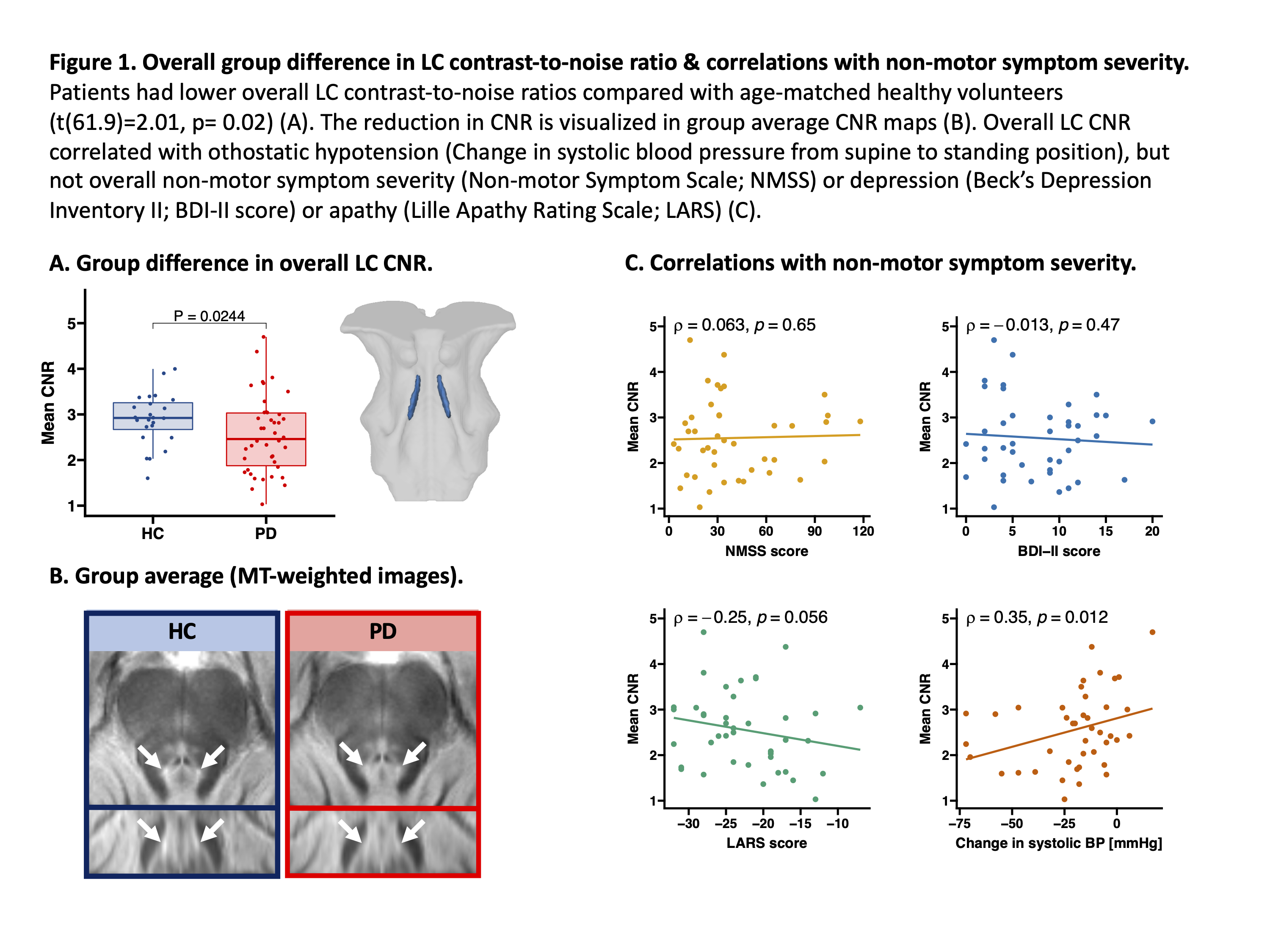
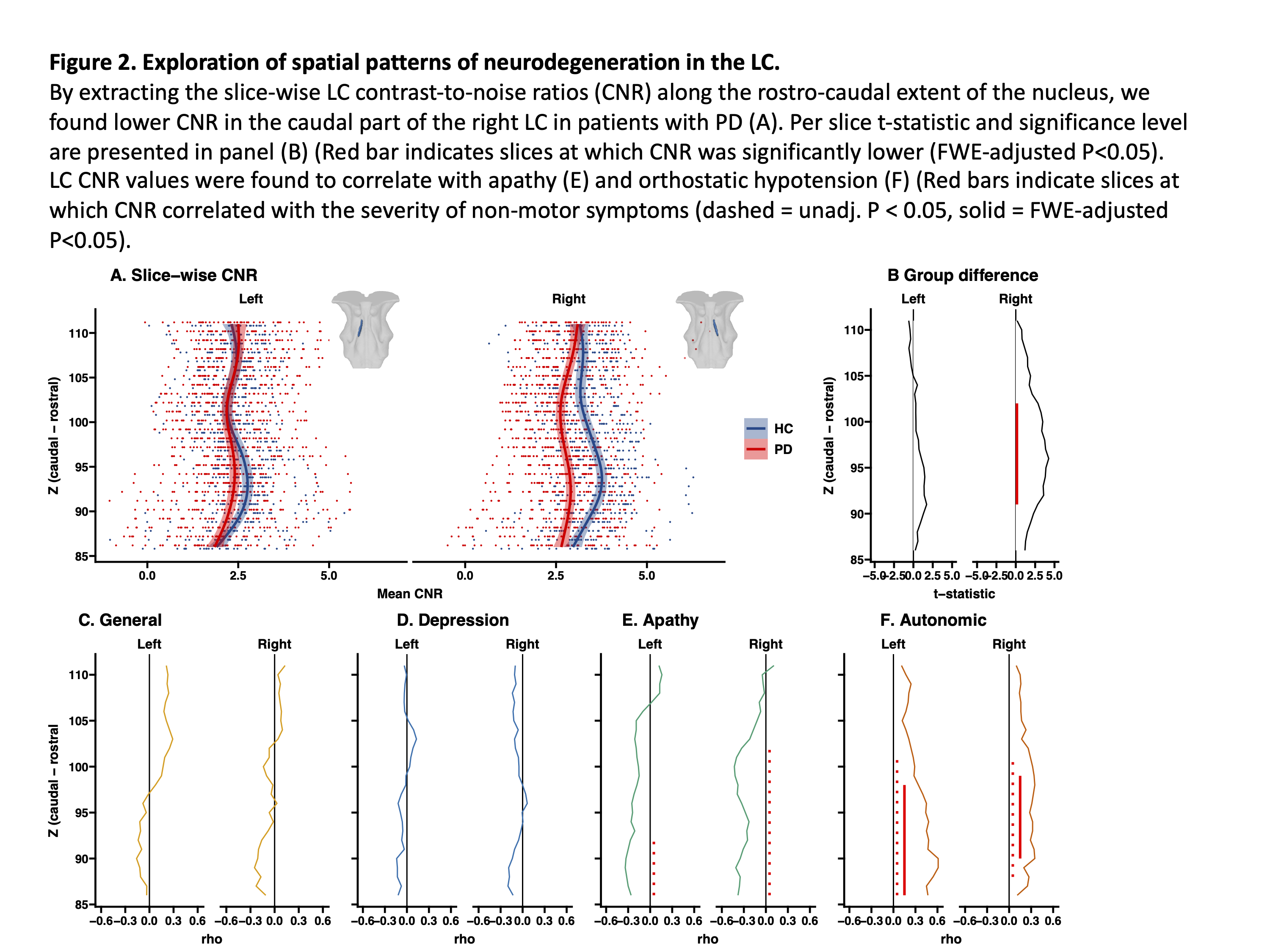
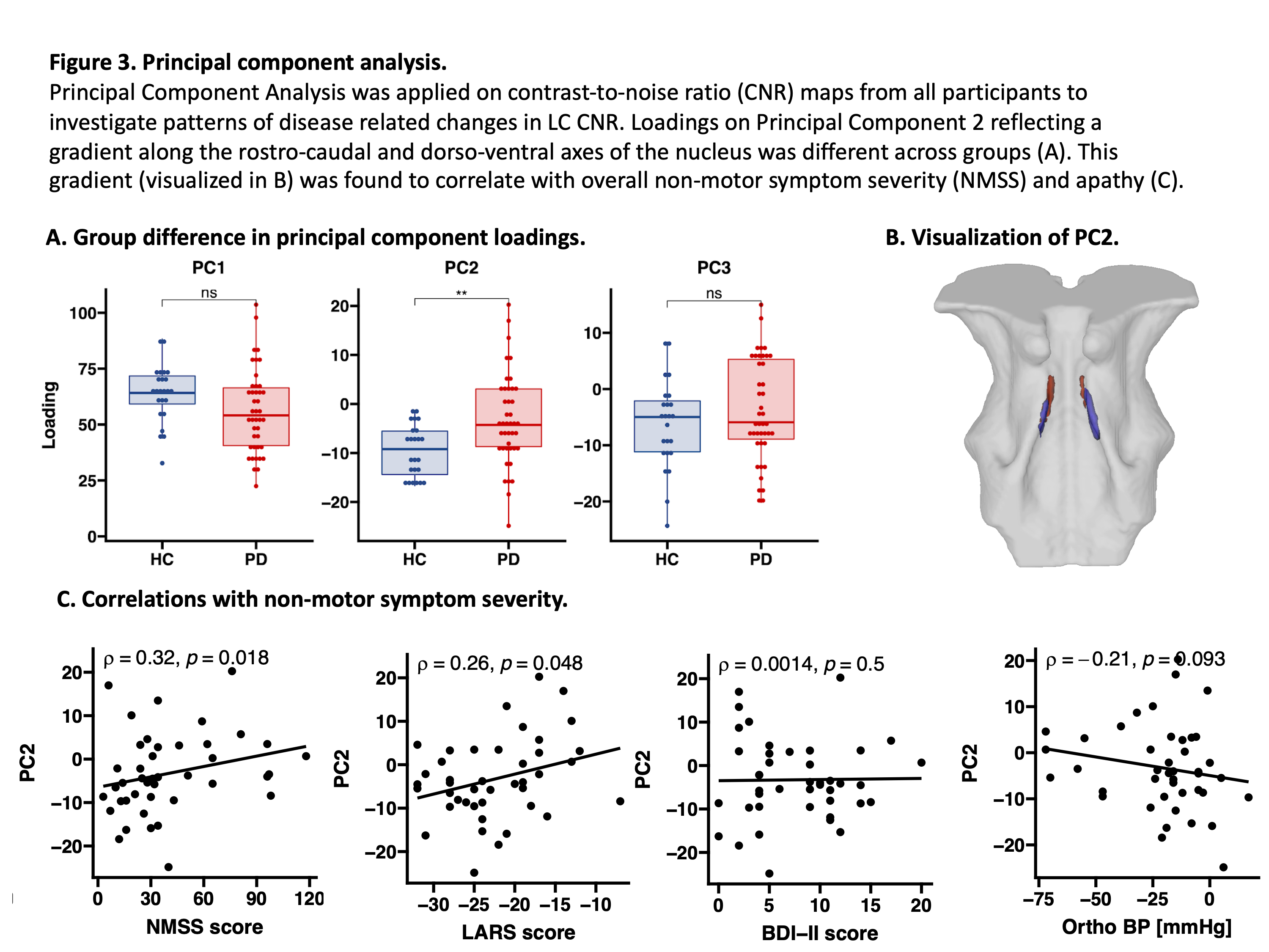
Results: Patients had a reduced signal caudally in the right LC relative to controls (Fig. 1 & 2). Accordingly, PCA revealed a rostro-caudal pattern of PD-related signal reduction with greater reduction in caudal parts (Fig. 3). Severity of apathy and orthostatic hypotension scaled positively with the reduction of LC signal in the PD group (Fig. 2E & F).
[figure1]
[figure2]
[figure3]
Conclusion: PD is associated with LC degeneration especially in the caudal part. The pattern of degeneration was found to correlate with measures non-motor symptom severity, particularly orthostatic hypotension.
References: [1] Vermeiren, Y. & De Deyn, P. P. Targeting the norepinephrinergic system in Parkinson’s disease and related disorders: The locus coeruleus story. Neurochemistry International 102, 22–32 (2017). [2] Betts, M. J. et al. Locus coeruleus imaging as a biomarker for noradrenergic dysfunction in neurodegenerative diseases. Brain 142, 2558–2571 (2019). [3] Ye, R. et al. An in vivo probabilistic atlas of the human locus coeruleus at ultra-high field. NeuroImage 225, 117487 (2021).
To cite this abstract in AMA style:
C. Madelung, D. Meder, S. Fuglsang, V. Boer, E. Petersen, A. Løkkegaard, H. Siebner. Does neurodegeneration of the noradrenergic locus coeruleus follow a spatial gradient in Parkinson’s disease? [abstract]. Mov Disord. 2021; 36 (suppl 1). https://www.mdsabstracts.org/abstract/does-neurodegeneration-of-the-noradrenergic-locus-coeruleus-follow-a-spatial-gradient-in-parkinsons-disease/. Accessed July 9, 2025.« Back to MDS Virtual Congress 2021
MDS Abstracts - https://www.mdsabstracts.org/abstract/does-neurodegeneration-of-the-noradrenergic-locus-coeruleus-follow-a-spatial-gradient-in-parkinsons-disease/


